Subject area:
Atom Probe Tomography and 3D-Nanoanalytics
Responsible employees:
Prof. Dr. Peter Felfer
Mehrpad Monajem (M. Sc.)
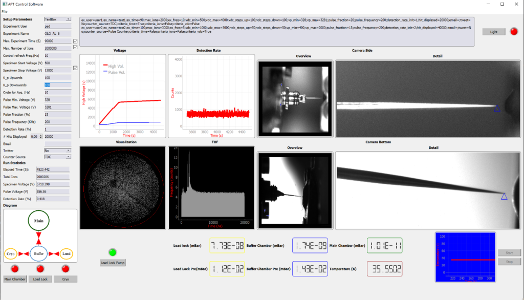
Figure: The user interface of the atom probe control system, including
data visualization. The valveand pump controls, as well as the camera view,
allow the user to insert and align the sample.
Atom Probe Tomography (APT) is a unique material characterisation method that can provide three-dimensional imaging and chemical composition at the atomic level [1]. The application of this technique has expanded beyond high-strength structural alloys and semiconductor materials to catalyst nanoparticles and organic metal frameworks [2]. APT can be used to image and analyze all chemicals in a solid sample without bias and with equal sensitivity, historically except for hydrogen. Many researchers use commercial atom probes for their projects. However, commercial atom probes used closed instrument hardware and locked-in control software. A titanium atom probe was built at the Material Science and Engineering Institute I to circumvent these issues. The design was centered on the achievement of low hydrogen vacuum levels [3]. Together with novel control schemes, this allows for the in-situ heat treatment and imaging of hydrogen in this atom probe. To achieve the highest data fidelity, low-level access to the detector data is required.
This project aims to image all atoms at interfaces in materials in 3D with low distortion to enable simulation of grain boundary behavior. To accomplish this, the project was divided into two steps. In the first step, control software will be implemented to control the atom probe experiments. This software must control the experiment, read the detector, and store the experiment data in Findable, Accessible, Interpretable, and Reusable (FAIR) format. Moreover, the software should save the data in raw format. A laser pulser and a digitizer based detector will also be added to this titanium atom probe in the near future, both of which require integration into the control software. This will allow us to image the interfaces with previously not achieved efficiency.
In the second step, the data must be calibrated and analyzed in order to achieve a low distortion 3D reconstruction of the sample. In this step, special reconstruction algorithms will be explored to improve the current methodology so that it can yield a more accurate picture at atomic scale.
[1] B. Gault, M.P. Moody, F. De Geuser, D. Haley, L.T. Stephenson, S.P. Ringer, Applied Physics Letters 95 (2009) 034103.
[2] Z. Ji, T. Li, O.M. Yaghi, Science 369 (2020) 674-680.
[3] P. Felfer, B. Ott, M. Monajem, V. Dalbauer, M. Heller, J. Josten, C. Macaulay, Microscopy and Microanalysis (2021) 1-9.